Home » Without Label » Tendon Diagram - Tendon anatomy diagram - Broadly considered, human muscle—like the muscles of all vertebrates—is often divided into striated muscle, smooth muscle, and cardiac muscle.
Tendon Diagram - Tendon anatomy diagram - Broadly considered, human muscle—like the muscles of all vertebrates—is often divided into striated muscle, smooth muscle, and cardiac muscle.
Tendon Diagram - Tendon anatomy diagram - Broadly considered, human muscle—like the muscles of all vertebrates—is often divided into striated muscle, smooth muscle, and cardiac muscle.. This hd wallpaper knee diagram tendons has viewed by 709 users. Tendons are the connection between bones and muscles tendon diagram. The hip itself is a ball and socket joint, much like the shoulder.the structures necessary to create this joint are the socket, the joint capsule, muscle, ligaments, and the neck. You can see a diagram of the achilles tendon below. The knee joint is a complex structure that involves bones.
The knee joint is a complex structure that involves bones. Tendons that attach parts of your head to your collarbone, breastbone, shoulder blades or bones in your back help you move your head and neck in different directions. Diagram of inside the body. Tendons are thick bands of tissue that connect muscles to bones. Broadly considered, human muscle—like the muscles of all vertebrates—is often divided into striated muscle, smooth muscle, and cardiac muscle.
8 Effective Achilles Tendon Treatments/Surgery in Scottsdale from www.drkosak.com By connecting our rigid bones to our powerful muscles, tendons allow us to move. Medical illustration showing carpal tunnel syndrome in the human wrist, and the surgical procedures associated with it. The changes in ligaments and tendons generally occur more slowly than adaptation in bone, because ligaments and tendons have less vascular supply. The fcu tendon is one of two tendons that bend the wrist. Both are made of collagen.ligaments connect one bone to another, while tendons connect muscle to bone. Your biceps tendons attach the biceps muscle to bones in the shoulder and in the elbow. Its muscle belly is in the forearm. The tendon runs down the back of your lower leg from the back of the knee to the heel.
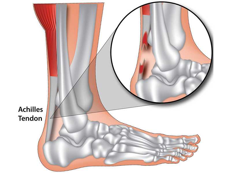
The achilles tendon is a tough band of fibrous tissue that connects the calf muscles to the heel bone (calcaneus).
Tendons are thick bands of tissue that connect muscles to bones. These muscles, acting via the tendon, cause plantar flexion of the foot at the ankle joint, and (except the soleus) flexion at the knee. Also allows the action of raising up onto toes. Again, our knowledge of how mechanical stimulus mediates ligament and tendon structure is more empirical and less. Foot anatomy diagram, foot joint diagram, foot sprain diagram, foot tendons and ligaments pain, leg tendon diagram, peroneal tendonitis, foot, foot anatomy diagram, foot joint diagram, foot sprain diagram, foot tendons and ligaments pain, leg tendon diagram, peroneal tendonitis. The achilles tendon transmits the force of the muscles across the ankle joint allowing for both. Your biceps tendons attach the biceps muscle to bones in the shoulder and in the elbow. The pubis, ischium, and ilium together constitute the pelvis while the thigh bone is the femur. The knee joint is a complex structure that involves bones. When the biceps contracts, it pulls the forearm up and rotates it outward. The tendon runs down the back of your lower leg from the back of the knee to the heel. Tendon diagram simple / 8.4c: Allows the action of raising the foot.
By connecting our rigid bones to our powerful muscles, tendons allow us to move. Bones in shoulder, ligaments of the shoulder joint, parts of the shoulder joint, shoulder anatomy, shoulder joints and muscles, shoulder structure anatomy, shoulder tendon anatomy, shoulder tendons ligaments, human muscles, bones in shoulder, ligaments of the shoulder joint, parts of. This hd wallpaper knee diagram tendons has viewed by 709 users. Tendons, located at each end of a muscle, attach muscle to bone. Human muscle system, the muscles of the human body that work the skeletal system, that are under voluntary control, and that are concerned with movement, posture, and balance.
Foot Tendons And Ligaments Diagram - Human Anatomy Body from www.anatomylibrary99.com The bones of the hip include the femur, the ilium, the ischium, and the pubis. The achilles tendon is also called the calcaneal tendon. Diagram depicting the bones, ligaments and muscles throughout the hand and fingers. Broadly considered, human muscle—like the muscles of all vertebrates—is often divided into striated muscle, smooth muscle, and cardiac muscle. The insertions of the tibialis posterior tendon are illustrated. 17 photos of the diagram of shoulder muscles and tendons. The pubis, ischium, and ilium together constitute the pelvis while the thigh bone is the femur. Tendon, tissue that attaches a muscle to other body parts, usually bones.
Tendons, located at each end of a muscle, attach muscle to bone.
Diagram of inside the body. Attaches the calf muscles to the calcaneus, most important muscles for running, jumping, walking etc. Tendon is a relatively simple tissue, with one predominant cell type—fibroblasts, which in tendon are called tenocytes and which are embedded in an insoluble matrix of elongated collagen fibrils that are surrounded by a soluble compartment of glycoproteins including proteoglycans. The insertions of the tibialis posterior tendon are illustrated. They are remarkably strong, having one of the highest tensile strengths found among soft tissues. You can see a diagram of the achilles tendon below. They are attached to the femur (thighbone), tibia (shinbone), and fibula (calf bone) by fibrous tissues called ligaments. The achilles tendon or heel cord, also known as the calcaneal tendon, is a tendon at the back of the lower leg, and is the thickest in the human body. Movement occurs when our muscles pull on our bones, relocating them. Allows the foot to be turned inward and also supports the arch of the foot. It serves to attach the plantaris, gastrocnemius (calf) and soleus muscles to the calcaneus (heel) bone. Tendons, located at each end of a muscle, attach muscle to bone. The achilles tendon is a tough band of fibrous tissue that connects the calf muscles to the heel bone (calcaneus).
The fleshy, thick part of the muscle is called its belly. Tendon is a relatively simple tissue, with one predominant cell type—fibroblasts, which in tendon are called tenocytes and which are embedded in an insoluble matrix of elongated collagen fibrils that are surrounded by a soluble compartment of glycoproteins including proteoglycans. The bones of the hip include the femur, the ilium, the ischium, and the pubis. Human muscle system, the muscles of the human body that work the skeletal system, that are under voluntary control, and that are concerned with movement, posture, and balance. One peroneal tendon attaches to the outer part of the midfoot, while the other tendon runs under the foot and attaches near the inside of the arch.
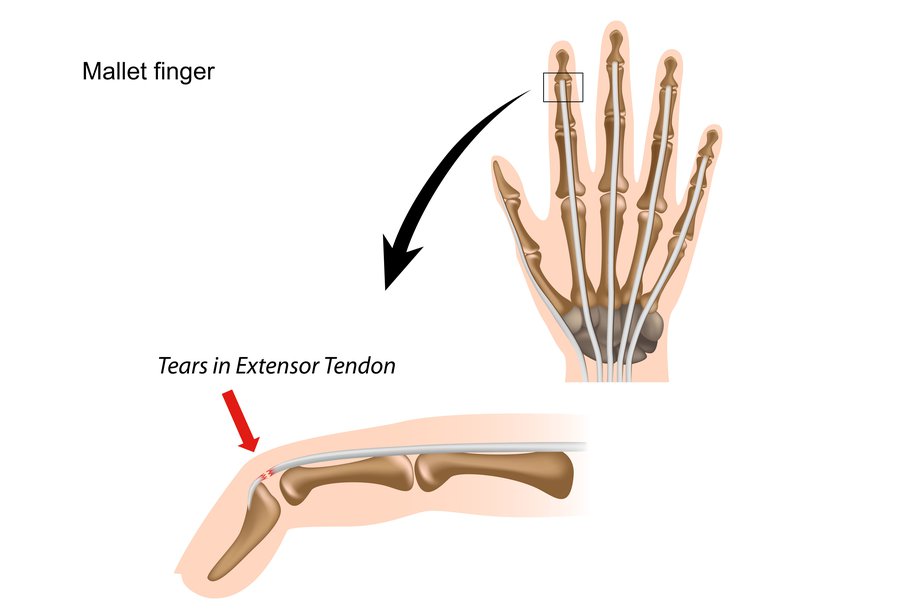
Mallet finger - NHS from assets.nhs.uk Tendons are found throughout the body, from the head and neck all the way down to the feet. Tendons are similar to ligaments; Movement occurs when our muscles pull on our bones, relocating them. Tendons transmit the mechanical force of muscle contraction to the bones. Human muscle system, the muscles of the human body that work the skeletal system, that are under voluntary control, and that are concerned with movement, posture, and balance. Broadly considered, human muscle—like the muscles of all vertebrates—is often divided into striated muscle, smooth muscle, and cardiac muscle. The pubis, ischium, and ilium together constitute the pelvis while the thigh bone is the femur. Tendons that attach parts of your head to your collarbone, breastbone, shoulder blades or bones in your back help you move your head and neck in different directions.
The achilles tendon or heel cord, also known as the calcaneal tendon, is a tendon at the back of the lower leg, and is the thickest in the human body.
The two peroneal tendons in the foot run side by side behind the outer ankle bone. The fleshy, thick part of the muscle is called its belly. Human muscle system, the muscles of the human body that work the skeletal system, that are under voluntary control, and that are concerned with movement, posture, and balance. Tendon diagram simple / 8.4c: The achilles tendon transmits the force of the muscles across the ankle joint allowing for both. Diagram of inside the body. Tendon, tissue that attaches a muscle to other body parts, usually bones. You can see a diagram of the achilles tendon below. The hip itself is a ball and socket joint, much like the shoulder.the structures necessary to create this joint are the socket, the joint capsule, muscle, ligaments, and the neck. 9 photos of the foot tendons and ligaments diagram. Foot anatomy diagram, foot joint diagram, foot sprain diagram, foot tendons and ligaments pain, leg tendon diagram. Movement occurs when our muscles pull on our bones, relocating them. Tendon diagrams and design force vectors.